INTRODUCTION
Methylation of DNA is a hereditable epigenetic marker. With the help of the DNA methyltransferase enzyme, a methyl group (CH3) is transferred to a DNA cytosine ring1. It controls duplex stability, imprinting, replication, repair, transcription, X-chromosome inactivation, and epigenetic memory2. The repressed chromatin of mammals showed higher levels of 5 mC. A low level of DNA methylation is harmful to human health3. It acts as a reliable predictor for disease progression4. Methylation of DNA is crucial for the regulation of gene expression and the development of cells. A specific region of the genome or the whole genome can be used for the methylation of DNA. The concentrations of LINE-1 (long-interspersed nuclear elements-1), 5 mC, and Alu (Arthrobacter luteus), can represent global DNA methylation. These markers could be used as a prior screening tool when compared to gene-specific regions.
Pb toxicity is mediated by increased levels of delta-aminolevulinic acid, decreased glutathione and activated Ca2+-dependent cysteine protease5. Lead-exposed workers experienced global (Alu and LINE-1) and gene-specific methylation changes due to lead-mediated genotoxicity and DNA damage6. Issah et al.7 found a weak association between LINE-1 methylation and Pb exposure in e-waste workers. Recent studies have found an altered 5 mC in Pb-exposed workers8,9. According to a literature review, it is necessary to consider the consequences of Pb exposure, such as dose and duration, when determining DNA methylation levels10.
B vitamins function as coenzymes and substrates for biological reactions. The synthesis of S-adenosylmethionine (SAM) requires B vitamins, including B6, B9, and B1211. The dietary folate is converted into 5, 10-methylene THF through dihydrofolate (DHF) and tetrahydrofolate (THF) using the enzymes dihydrofolate reductase, serine hydroxylmethyltransferase, and vitamin B6. Methylenetetrahydrofolate reductase (MTHFR) and vitamin B12 act as cofactors to create methylcobalamin by converting 5, 10-methyleneTHF to 5-methyleneTHF. Vitamins B9 and B12 are essential for the formation of methionine from homocysteine. Methionine is transformed into SAM using the enzyme methyl-Adenosyl transferase. The released methyl (CH3) groups are necessary for DNA, RNA, hormones, neurotransmitters, lipids, and protein biomolecules12. Mandaviya et al.13 revealed that low levels of B vitamins affect DNA methylation. Brunaud et al.14 reported that deficiencies of vitamin B12 and methionine synthase are strong predictors of DNA methylation14. The diet with low levels of B9 and B12 observed indistinct genomic methylation of DNA15. Buyuksekerci et al.16 evaluated the status of vitamin B9 and B12 in Pb-exposed workers and observed considerably reduced levels when compared to unexposed workers. As per the literature review, there is a knowledge gap regarding the impact of B vitamin deficiencies on DNA methylation among Pb-exposed workers.
Lifestyle factors are adaptive behaviors that have a consequence on health. In earlier investigations, gender, age, and smoking behaviors were associated with Pb exposure17. Previous research has indicated also that sedentary habits, advanced age, and inferior socioeconomic status were associated with vitamin B12 deficiency18. Furthermore, Thuesen et al.19 found that a poor diet, smoking, excessive alcohol and coffee drinking were all associated with vitamin B9 deficiency. Personalized nutrition, alcohol intake, ageing, and vitamin malabsorption modify epigenetic expression20. Based on the above, lifestyle factors influence the status of Pb exposure, B vitamins and gene expressions, and hence there is a need to ascertain how lifestyle factors may affect DNA methylation in Pb-exposed workers. In relation to global DNA methylation, the effects of Pb exposure consequences (dose and time), B-vitamin deficits and lifestyle variables have not been examined. The current research aimed to evaluate the association of Pb-exposure and global DNA methylation by considering lifestyle variables and B vitamin deficiency among lead-recycling workers.
METHODS
This cross-sectional study took place in 2021–2022 in India, among a convenience sample of 164 male workers exposed occupationally to Pb. There were no female workers in this recycling process. Pb-exposed workers who were willing to participate and were aged ≥18 years were included in the study. The sample size was selected based on the prevalence of vitamin B12 deficiency (p=0.65) among professionals21 with 90% CI and 10% of relative precision. The sample obtained from this calculation is 145. Subjects who were taking oral or injectable B vitamin supplementation continuously for >15 days during the previous one year, had known cases of cardiovascular diseases, were diabetic with Metformin treatment and recently donated or received blood, were excluded from the study.
Questionnaire
A pre-structured questionnaire was used to collect individual data on age, occupational exposure details, marital status (married, unmarried), habits of smoking, alcohol consumption (yes, no), chewing of tobacco products (yes, no), and diet type (vegetarian, non-vegetarian) among workers. The worker BMI (kg/m2) was calculated. The definition of smoking status and alcohol consumption status (never drinking, currently drinking) among study subjects followed the guidelines of Sanford et al.22 and Mulia et al.23.
Sample collection
For each worker, 4 mL of whole blood was collected, and the sample was distributed into two portions. One portion (1 mL) of sample was transferred into a dark green capped heparinized tube (Lab Tech Disposables, India) for BLL analysis. Another portion of whole blood sample (3 mL) was transferred into a red capped tube with an easy clot activator (Lab Tech Disposables, India). Centrifuging was used to separate the serum for 10 min at 4000 rpm and stored at 4°C. B vitamins (B6, B9, and B12) and DNA methylation tests were performed on the separated serum samples.
Blood lead levels (BLLs)
The lead level in blood samples was estimated using the Hasania et al.24 method. A volume of 0.5 mL of heparinized whole blood was combined with 2 mL of 69% HNO3 and 1.0 mL of 30% hydrogen peroxide (H2O2) for the protocol’s digestion. It was digested by using ETHOS-D milestone microwave system. The digested sample was filtered using a membrane filter assembly, and the final volume of 7 mL was made using triple-distilled water. The level of Pb in the blood was quantified using inductively coupled plasma-optical emission spectrometry (Model iCAP 7000 Series, Thermo Scientific, USA). The analysis of the recovery rate, which was used as a quality control tool, showed a 98% recovery rate. The LOD and LOQ of the method were calculated using Pb concentration (0–100 µg/L) versus intensity (cycles/s). The LOD and LOQ of the method were found to be 7.0 and 21 µg/L, respectively. The BLLs in the samples were calculated using the dilution factor and converted to µg/dL.
Vitamins B6 (pyridoxine), B9 (folic acid) and B12 (cobalamin)
The level of vitamin B6 was quantified using the ELISA method (BT Lab, cat. no. E1539Hu, China). In this method, the vitamin B6 in the sample binds to the human vitamin B6 antibody that is treated with biotinylated human vitamin B6 antibody and streptavidin – HRP. After incubation, the substrate solution was added to produce color. An acidic solution was used to terminate the reaction. Sample and standard transmittance were detected by using a Lisa Scan EM microplate reader at 450 nm (Transasia Biomedical Limited, India). The increase in transmittance relates to vitamin B6 levels in the sample. The LOD of the protocol is 2.51 pmol/L, and the detection range is 5–1500 pmol/L. The normal and deficient distribution of vitamin B6 was evaluated using the Obol et al.25 guidelines.
The level of vitamin B9 was quantified using the ELISA method (BT Lab, cat. no. E3930Hu, China). In this technique, the vitamin B6 in the sample binds to the human vitamin B9 antibody that is treated with biotinylated human vitamin B6 antibody and streptavidin – HRP. After incubation, the substrate solution was added to produce color. An acidic solution was used to terminate the reaction. Sample and standard transmittance were detected by using a Lisa Scan EM microplate reader at 450 nm (Transasia Biomedical Limited, India). The increase in transmittance relates to vitamin B9 levels in the sample. The LOD of the method is 0.03 ng/mL, and the measured range is 0.05–20 ng/mL. The normal and deficient frequency distributions of B9 were evaluated using WHO-recommended limits26.
The level of vitamin B12 was quantified using the ELISA (CAL Biotech, cat. no. VB369B, USA). In this procedure, the vitamin B12 in the sample binds to the human vitamin B12 antibody that is treated with biotinylated human vitamin B6 antibody and streptavidin – HRP. After incubation, the substrate solution was added to produce color. An acidic solution was used to terminate the reaction. Sample and standard transmittance were detected by using a Lisa Scan EM microplate reader at 450 nm (Transasia Biomedical Limited, India). The increase in transmittance relates to vitamin B12 levels in the sample. The limit of delectation of the protocol is 41 pg/mL, and the quantification range is 0–2000 pg/mL. The normal and deficient frequency distributions of vitamin B12 was evaluated using WHO-recommended limits26.
DNA isolation and global DNA methylation (5 mC)
The Fit Amp TM protocol was used to isolate serum DNA (Epigentek, Cat. P-1004, USA). This protocol allows DNA quantification between 0.1 and 2 µg, with an optimal range of 1 ng to 1 µg. The yield and purity of the isolated DNA were measured at a 260/280 nm ratio using a Nanodrop Spectrophotometer (Thermo ScientificTM Nano DropTM). The ELISA protocol was employed to measure the global methylation of the isolated DNA (Epigentek, cat. no. P 1030, USA). The methylated fraction of DNA is quantified using a Lisa Scan EM microplate reader at 450 nm (Transasia Biomedical Limited, India). The proportion of DNA methylation is relative to the intensity of absorbance measured. The detection limit of the method is 0.05% methylated DNA from 100 ng of input DNA. The percent of 5 mC in total DNA was calculated.
Statistical analysis
Data analysis was done using SPSS version 23 software (IBM Corp., Armonk, NY, USA). The DNA methylation among workers was found to be a non-normal distribution when Kolmogorov-Smirnov and Shapiro-Wilk tests were performed. The data were expressed as frequencies and percentages, and mean rank of log transformed 5 mC. Mann-Whitney U and Kruskal-Wallis H tests were used to compare categorical variables on DNA methylation. The correlation between DNA methylation (5 mC), and demographic, lifestyle factors, B vitamins, and BLLs were assessed using the Spearman correlation coefficient (r). We used a stepwise regression model to assess the relationship between DNA methylation (5 mC) and potential predictors. BLLs, experience, B vitamins, and lifestyle factors (age, BMI, smoking, alcohol consumption, tobacco chewing, marital status, and diet) were used as potential covariates. In this model, the covariates were added or removed based on the test statistics of the estimated coefficients with the F-test. A p<0.05 was used for statistical significance.
RESULTS
Table 1 displays the demographic information of Pb-exposed workers. The workers with the highest percentage were aged 21–30 years, while the workers with the lowest percentage were aged <20 years. The BMI values indicated that 36% of subjects are within the normal range, 18.3% were overweight, and 39% were obese, and 6.7% were underweight. The highest proportion of workers had <5 years of exposure, 67.1% of the study participants were married, 32.9% consumed alcohol, 22% chewed tobacco, 12% were current smokers, and the majority had mixed diets of vegetarian and non-vegetarian foods. Among the workers, 39% had a vitamin B12 deficiency, 28.7% a vitamin B9 deficiency, and 57.9% had a vitamin B6 deficiency.
Table 1
Demographic details of lead-recycling workers, a cross-sectional study, India 2022 (N=164)
Table 2 shows the results of the variables that affect global DNA methylation (5 mC) among Pb-exposed workers. The variables like age, BMI, smoking, alcohol consumption, diet and B vitamins status were compared using non-parametric tests. A Mann-Whitney U test was employed to compare the effects of variables of experience (≤5 and >5 years), marital status (married, unmarried), alcohol consumption (yes, no), tobacco chewing (yes, no), diet (vegetarian, non-vegetarian), vitamin B6 (normal, deficient), vitamin B9 (normal, deficient), and vitamin B12 (normal, deficient) on global DNA methylation among Pb-exposed workers. The results showed that experience and vitamin B9 deficiency significantly reduced 5 mC. The impact of categorical variables such as age, and BMI on 5 mC was assessed using the Kruskal-Wallis H test. The variables age and BMI were found to have no significant impact on 5 mC.
Table 2
The effect of variables on global DNA methylation (5 mC) among lead-recycling workers, a cross-sectional study, India, 2022 (N=164)
| Variables | n | % | Mean rank | p |
|---|---|---|---|---|
| Age (years) | ||||
| ≤20 | 10 | 6.1 | 109.25 | 0.173 |
| 21–30 | 73 | 44.5 | 81.28 | |
| 31–40 | 50 | 30.5 | 85.51 | |
| ≥41 | 31 | 18.9 | 71.89 | |
| BMI (kg/m2) | ||||
| <18.5 (underweight) | 11 | 6.7 | 105.50 | 0.287 |
| 18.5–22.9 (normal) | 59 | 36 | 85.68 | |
| 23–24.9 (overweight) | 30 | 18.3 | 77.43 | |
| ≥25 (obese) | 64 | 39 | 77.99 | |
| Experience (years) | ||||
| ≤5 | 100 | 61 | 89.80 | 0.014* |
| >5 | 64 | 39 | 71.10 | |
| Marital status | ||||
| Married | 110 | 67.1 | 77.39 | 0.049* |
| Unmarried | 54 | 32.9 | 92.91 | |
| Alcohol consumption | ||||
| No | 110 | 67.1 | 82.42 | 0.976 |
| Yes | 54 | 32.9 | 82.66 | |
| Tobacco chewing | ||||
| No | 128 | 78 | 84.68 | 0.268 |
| Yes | 36 | 22 | 74.76 | |
| Smoking habits | ||||
| Non-smoker | 131 | 79.9 | 82.79 | 0.974 |
| Current smoker | 20 | 12 | 80.23 | |
| Ex-smoker | 13 | 7.9 | 83.04 | |
| Diet | ||||
| Vegetarian | 15 | 9.1 | 71.07 | 0.327 |
| Non-vegetarian | 149 | 90.9 | 83.65 | |
| Vitamin B6 (pyridoxine) | ||||
| Normal (≥235 pmol/L) | 69 | 42.1 | 87.96 | 0.209 |
| Deficient (<235 pmol/L) | 95 | 57.9 | 78.54 | |
| Vitamin B9 (folic acid) | ||||
| Normal (≥3 ng/mL) | 117 | 71.3 | 87.28 | 0.042* |
| Deficient (<3 ng/mL) | 47 | 28.7 | 70.61 | |
| Vitamin B12 (cobalamin) | ||||
| Normal (≥203 pg/mL) | 100 | 61 | 82.57 | 0.983 |
| Deficient (<203 pg/mL) | 64 | 39 | 82.40 |
Table 3 shows the relationship between global DNA methylation and BLLs, lifestyle factors and B-vitamins among subjects, based on the Spearman correlation coefficient (r). Global DNA methylation was found to have a negative association with age, BMI, experience, smoking, tobacco chewing, BLLs, and vitamin B9. A significant association was found with marital status and job experience.
Table 3
Spearman correlation coefficient (r) between DNA methylation (5 mC) and lifestyle variables, B vitamins and lead-exposure, among lead-recycling workers, a cross-sectional study, India, 2022 (N=164)
| Variables | Spearman correlation coefficient (r) |
|---|---|
| Age (years) | -0.125 |
| Body mass index (kg/m2) | -0.108 |
| Marital status | -0.154* |
| Tobacco chewing | -0.087 |
| Smoking | -0.011 |
| Alcohol consumption | 0.002 |
| Diet | 0.077 |
| Experience (years) | -0.168* |
| Vitamin B6 | 0.066 |
| Vitamin B12 | 0.086 |
| Vitamin B9 | -0.018 |
| BLLs | -0.109 |
Table 4 shows the stepwise multiple linear regression analysis of variables that affect DNA methylation among Pb-exposed workers. In this model, the 5 mC is used as the dependent variable, and the variables age, BMI, experience, diet, smoking, alcohol consumption, tobacco chewing, BLLs, and B vitamins (B6, B9, and B12) were used as independent variables. Experience (β= -0.283; p=0.001), increased BLLs (β= -0.195; p=0.010), tobacco chewing (β= -0.184; p=0.014), and vitamin B9 deficiency (β= -0.157; p=0.037) were all significantly related to global DNA methylation. The model explains 12.7% of the variation in global DNA methylation. Age, BMI, diet, smoking, alcohol consumption, and B vitamins (B6 and B12) did not significantly affect DNA methylation.
Table 4
Multiple regression analysis of variables that affect global DNA methylation (5 mC) among lead-recycling workers, a cross-sectional study, India, 2022 (N=164)
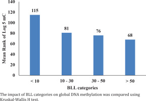
Figure 1 displays mean rank of DNA methylation in Pb-exposed workers according to their BLLs categories. The BLLs categories were 8.5%, 47.6%, 34.8%, and 9.1% for <10, 10–30, 30–50, and >50 µg/dL, respectively. The impact of BLLs categories on global DNA methylation was compared using Kruskal-Wallis H test. The mean ranks of DNA methylation were found to decrease (p=0.043) considerably with increase in BLLs.
DISCUSSION
The impact of Pb-exposure, B vitamin deficiencies, and lifestyle variables on global DNA methylation among Pb-exposed workers was examined. BLLs measurement was used as the internal dose of the body. In the present investigation, we estimated BLLs using the ICP-OES method since it was considered a better-quality research tool for biology and had easy handling, better sensitivity, support considerable amounts of dissolved solids, and sufficient analytical competence27.
The total genomic methylation was measured by using the levels of 5 mC, LINE-1, and Alu. In the present study, we determined 5 mC concentrations for DNA methylation using the ELISA protocol. This technique was quick, simple, and less expensive, required a small amount of DNA input, and could detect considerable altered DNA methylation with permissible large number of sample analyses28. According to the de Araújo et al.9 study, Pb-exposed workers with BLLs ranging from 2.6 to 48 µg/dL and an average duration of 3 years experienced a 3% of 5 mC. Devoz et al.8 found 2.8% of 5 mC with BLLs ranging from 1.8 to 48 µg/dL and a mean exposure time of 2.8 years. This study found 1.7% 5 mC with BLLs ranging from 3 to 79 µg/dL and a mean exposure time of 5.4 years. Comparing this study to previous studies, we found a reduced 5 mC, which brought a wider range of BLLs and a longer exposure time. Furthermore, this study also investigated the dose-effect association between BLLs and 5 mC. The level of 5 mC was considerably reduced in BLL for categories 30–50 and >50 µg/dL when compared to <10 µg/dL. The present investigation found a negative and considerable relationship between BLLs and 5 mC. Previous studies also noted a similar association between these parameters8,9.
We employed B vitamin assessments in workers to evaluate the normal and deficient status of B vitamins. We used immunoassay methods to estimate B vitamins in the serum samples of the workers. These assays worked on the principle of antibody and antigen reactions. These methods are capable of detecting low concentrations and do not require arduous extraction and clean-up procedures. Sivaprasad et al.29 reported 35% vitamin B12 and 12% vitamin B9 deficiencies in a community-based investigation. This study found that the vitamin B6, B9, and B12 deficiencies were 39%, 28.5%, and 57. 9%, respectively. Adolescents in India exhibited 39.8% vitamin B9 deficiency30. Obol et al.25 found that 42% of control participants had vitamin B6 deficiency. In our study, vitamin B6, B9, and B12 deficiencies were more prevalent than in community-based research studies. The production of DNA methylation depends on methyl nutrients such as pyridoxine, folate, and cobalamin. These substances also function as cofactors and substrates in one-carbon metabolism3. Since methylation of DNA capacity depends on the availability of methyl nutrients, we evaluated the impact of methyl nutrient deficiencies (B6, B9, and B12) on global DNA methylation among Pb-exposed workers.
Vitamin B9 and B12 deficiencies contribute to decreased global DNA methylation. According to Yadav et al.31, folate deficiency was a strong predictor of DNA methylation in a population residing in highly polluted areas. We found reduced DNA methylation in deficiencies of vitamins B6, B9, and B12, and a significant decrease was noted in vitamin B9 deficiency. Mandaviya et al.14 reported a negative relationship between B vitamins (B9 and B12) and DNA methylation. The present investigation also found a similar association between these parameters. Recent research has suggested that the supplementation of methyl nutrients protects from methylation loss and suppresses disease development32.
We also investigated how lifestyle factors affected DNA methylation. We specifically examined the possible confounding variables like age, body mass index, smoking, use of alcohol, chewing tobacco, and diet. We found that as workers get older, their DNA methylation decreases. This was implied due to the continuous cell division process. We also note that there was no noteworthy relationship between BMI categories and 5 mC. According to Zong et al.33, a current smoker alters epigenetics by activating enzymes involved in epigenetic modification and smoking-mediated inflammation. In the present study, it was found that there was decreased methylation of DNA in current smokers compared to non-smokers and ex-smokers. We also investigated the effect of tobacco chewing habits on DNA methylation and found a significant association.
According to a recent study, alcohol use reduces the body’s ability to absorb dietary folate, and in turn alters DNA methylation34. In the present study, alcohol use was found to reduce global DNA methylation, but it was not significant because the workers were occasional drinkers. A vegetarian diet contains lower levels of vitamin B12, which raises homocysteine levels and decreases methylation status35. Our research found no considerable difference between vegetarians and non-vegetarians with respect to 5 mC concentration. Most of the workers ate non-vegetarian food. According to Sbarra et al.36, DNA methylation has an inverse relationship with marital separation and divorce36. The current study found a significant relationship between DNA methylation and marital status (married, unmarried) among workers.
Limitations
The current study was conducted on a limited sample of Pbexposed workers. The study encountered several challenges, including residual confounding, non-casual design, lack of formal interactions and no female workers, and restricted generalized skills to do DNA methylation subgroup analysis. It is advised that large sample sizes and the previously specified information be included in future research.


